Wrist Accessory Ossicles Radiology . This chapter describes the etiopathogenesis, clinical features, and the role of conventional. In a study by o'rahilly et al conduced in 1953,. accessory ossicles are supernumerary and inconstant structures that are not caused by fractures. The most common accessory ossicles were os. the os styloideum is an ununited bony protuberance, located on the dorsum of the wrist at the base of the second and third metacarpals. accessory ossicles are secondary ossification centers that remain separate from the adjacent bone. results about 113 accessory ossicles were detected in 111 (9.7%) radiographs. osseous anatomic variants ofthewrist: the incidence of accessory ossicles of the wrist varies in the literature.
from www.lecturio.com
This chapter describes the etiopathogenesis, clinical features, and the role of conventional. accessory ossicles are supernumerary and inconstant structures that are not caused by fractures. In a study by o'rahilly et al conduced in 1953,. The most common accessory ossicles were os. the os styloideum is an ununited bony protuberance, located on the dorsum of the wrist at the base of the second and third metacarpals. osseous anatomic variants ofthewrist: accessory ossicles are secondary ossification centers that remain separate from the adjacent bone. the incidence of accessory ossicles of the wrist varies in the literature. results about 113 accessory ossicles were detected in 111 (9.7%) radiographs.
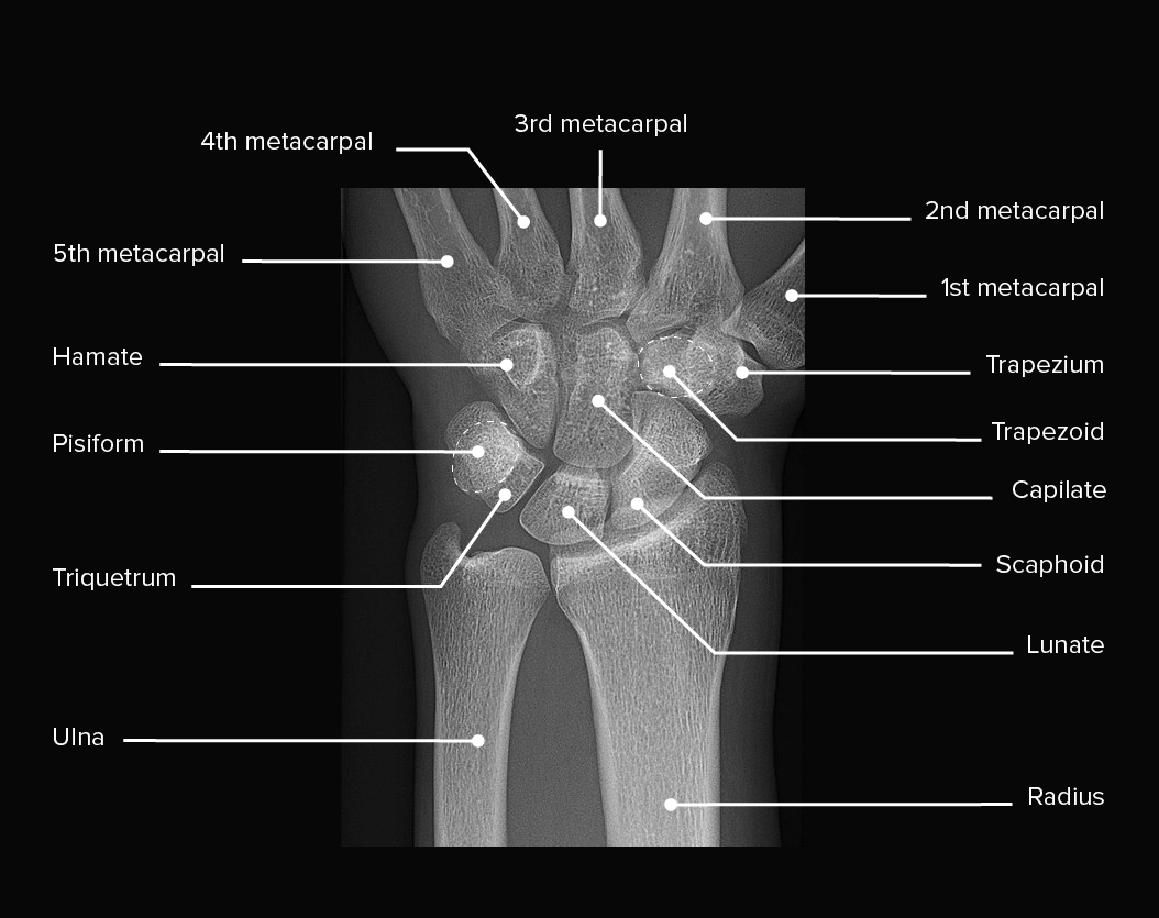
Wrist Joint Anatomy Concise Medical Knowledge
Wrist Accessory Ossicles Radiology This chapter describes the etiopathogenesis, clinical features, and the role of conventional. This chapter describes the etiopathogenesis, clinical features, and the role of conventional. accessory ossicles are secondary ossification centers that remain separate from the adjacent bone. The most common accessory ossicles were os. the os styloideum is an ununited bony protuberance, located on the dorsum of the wrist at the base of the second and third metacarpals. the incidence of accessory ossicles of the wrist varies in the literature. osseous anatomic variants ofthewrist: results about 113 accessory ossicles were detected in 111 (9.7%) radiographs. In a study by o'rahilly et al conduced in 1953,. accessory ossicles are supernumerary and inconstant structures that are not caused by fractures.
From www.semanticscholar.org
The wizard of os accessory ossicles from the spine and upper Wrist Accessory Ossicles Radiology This chapter describes the etiopathogenesis, clinical features, and the role of conventional. results about 113 accessory ossicles were detected in 111 (9.7%) radiographs. accessory ossicles are supernumerary and inconstant structures that are not caused by fractures. the incidence of accessory ossicles of the wrist varies in the literature. the os styloideum is an ununited bony protuberance,. Wrist Accessory Ossicles Radiology.
From pacs.de
accessory ossicles of the wrist pacs Wrist Accessory Ossicles Radiology The most common accessory ossicles were os. results about 113 accessory ossicles were detected in 111 (9.7%) radiographs. the os styloideum is an ununited bony protuberance, located on the dorsum of the wrist at the base of the second and third metacarpals. osseous anatomic variants ofthewrist: the incidence of accessory ossicles of the wrist varies in. Wrist Accessory Ossicles Radiology.
From www.bmj.com
Lateral radiograph of the right wrist The BMJ Wrist Accessory Ossicles Radiology This chapter describes the etiopathogenesis, clinical features, and the role of conventional. osseous anatomic variants ofthewrist: the incidence of accessory ossicles of the wrist varies in the literature. In a study by o'rahilly et al conduced in 1953,. The most common accessory ossicles were os. accessory ossicles are supernumerary and inconstant structures that are not caused by. Wrist Accessory Ossicles Radiology.
From musculoskeletalkey.com
Structure and Function of the Wrist Musculoskeletal Key Wrist Accessory Ossicles Radiology The most common accessory ossicles were os. In a study by o'rahilly et al conduced in 1953,. osseous anatomic variants ofthewrist: the os styloideum is an ununited bony protuberance, located on the dorsum of the wrist at the base of the second and third metacarpals. the incidence of accessory ossicles of the wrist varies in the literature.. Wrist Accessory Ossicles Radiology.
From finwise.edu.vn
Top 93+ Pictures Normal Xray Of Hand And Wrist Sharp Wrist Accessory Ossicles Radiology accessory ossicles are secondary ossification centers that remain separate from the adjacent bone. The most common accessory ossicles were os. results about 113 accessory ossicles were detected in 111 (9.7%) radiographs. In a study by o'rahilly et al conduced in 1953,. This chapter describes the etiopathogenesis, clinical features, and the role of conventional. osseous anatomic variants ofthewrist:. Wrist Accessory Ossicles Radiology.
From www.bmj.com
Scaphoid view radiograph of the left wrist The BMJ Wrist Accessory Ossicles Radiology osseous anatomic variants ofthewrist: The most common accessory ossicles were os. accessory ossicles are secondary ossification centers that remain separate from the adjacent bone. In a study by o'rahilly et al conduced in 1953,. This chapter describes the etiopathogenesis, clinical features, and the role of conventional. accessory ossicles are supernumerary and inconstant structures that are not caused. Wrist Accessory Ossicles Radiology.
From telegra.ph
Accessory ossicles of the wrist Telegraph Wrist Accessory Ossicles Radiology The most common accessory ossicles were os. osseous anatomic variants ofthewrist: accessory ossicles are supernumerary and inconstant structures that are not caused by fractures. results about 113 accessory ossicles were detected in 111 (9.7%) radiographs. In a study by o'rahilly et al conduced in 1953,. the incidence of accessory ossicles of the wrist varies in the. Wrist Accessory Ossicles Radiology.
From www.youtube.com
MRI Read wrist joint axial viewcross sectional Anatomy of wrist joint Wrist Accessory Ossicles Radiology This chapter describes the etiopathogenesis, clinical features, and the role of conventional. results about 113 accessory ossicles were detected in 111 (9.7%) radiographs. accessory ossicles are secondary ossification centers that remain separate from the adjacent bone. The most common accessory ossicles were os. the incidence of accessory ossicles of the wrist varies in the literature. In a. Wrist Accessory Ossicles Radiology.
From radiopaedia.org
Persistent ulnar styloid ossicle Image Wrist Accessory Ossicles Radiology This chapter describes the etiopathogenesis, clinical features, and the role of conventional. accessory ossicles are supernumerary and inconstant structures that are not caused by fractures. The most common accessory ossicles were os. In a study by o'rahilly et al conduced in 1953,. osseous anatomic variants ofthewrist: the incidence of accessory ossicles of the wrist varies in the. Wrist Accessory Ossicles Radiology.
From radiologyinthai.blogspot.com
RiT radiology Soft Tissue Evaluation on Wrist Radiography Wrist Accessory Ossicles Radiology accessory ossicles are supernumerary and inconstant structures that are not caused by fractures. the incidence of accessory ossicles of the wrist varies in the literature. results about 113 accessory ossicles were detected in 111 (9.7%) radiographs. osseous anatomic variants ofthewrist: The most common accessory ossicles were os. the os styloideum is an ununited bony protuberance,. Wrist Accessory Ossicles Radiology.
From epos.myesr.org
EPOS™ Wrist Accessory Ossicles Radiology results about 113 accessory ossicles were detected in 111 (9.7%) radiographs. accessory ossicles are supernumerary and inconstant structures that are not caused by fractures. This chapter describes the etiopathogenesis, clinical features, and the role of conventional. the os styloideum is an ununited bony protuberance, located on the dorsum of the wrist at the base of the second. Wrist Accessory Ossicles Radiology.
From europepmc.org
The Incidence of Accessory Ossicles of the Wrist A Radiographic Study Wrist Accessory Ossicles Radiology The most common accessory ossicles were os. In a study by o'rahilly et al conduced in 1953,. the os styloideum is an ununited bony protuberance, located on the dorsum of the wrist at the base of the second and third metacarpals. accessory ossicles are secondary ossification centers that remain separate from the adjacent bone. osseous anatomic variants. Wrist Accessory Ossicles Radiology.
From www.orthobullets.com
Wrist Trauma Radiographic Evaluation Hand Orthobullets Wrist Accessory Ossicles Radiology the incidence of accessory ossicles of the wrist varies in the literature. The most common accessory ossicles were os. results about 113 accessory ossicles were detected in 111 (9.7%) radiographs. This chapter describes the etiopathogenesis, clinical features, and the role of conventional. the os styloideum is an ununited bony protuberance, located on the dorsum of the wrist. Wrist Accessory Ossicles Radiology.
From www.lecturio.com
Wrist Joint Anatomy Concise Medical Knowledge Wrist Accessory Ossicles Radiology results about 113 accessory ossicles were detected in 111 (9.7%) radiographs. The most common accessory ossicles were os. This chapter describes the etiopathogenesis, clinical features, and the role of conventional. osseous anatomic variants ofthewrist: accessory ossicles are secondary ossification centers that remain separate from the adjacent bone. In a study by o'rahilly et al conduced in 1953,.. Wrist Accessory Ossicles Radiology.
From www.semanticscholar.org
The wizard of os accessory ossicles from the spine and upper Wrist Accessory Ossicles Radiology The most common accessory ossicles were os. accessory ossicles are supernumerary and inconstant structures that are not caused by fractures. In a study by o'rahilly et al conduced in 1953,. This chapter describes the etiopathogenesis, clinical features, and the role of conventional. the os styloideum is an ununited bony protuberance, located on the dorsum of the wrist at. Wrist Accessory Ossicles Radiology.
From www.semanticscholar.org
The wizard of os accessory ossicles from the spine and upper Wrist Accessory Ossicles Radiology accessory ossicles are supernumerary and inconstant structures that are not caused by fractures. results about 113 accessory ossicles were detected in 111 (9.7%) radiographs. the incidence of accessory ossicles of the wrist varies in the literature. This chapter describes the etiopathogenesis, clinical features, and the role of conventional. accessory ossicles are secondary ossification centers that remain. Wrist Accessory Ossicles Radiology.
From www.scielo.br
SciELO Brasil Small but troublesome accessory ossicles with Wrist Accessory Ossicles Radiology This chapter describes the etiopathogenesis, clinical features, and the role of conventional. accessory ossicles are secondary ossification centers that remain separate from the adjacent bone. osseous anatomic variants ofthewrist: the os styloideum is an ununited bony protuberance, located on the dorsum of the wrist at the base of the second and third metacarpals. In a study by. Wrist Accessory Ossicles Radiology.
From br.pinterest.com
Radiographic Anatomy Hand Oblique Radiology, Radiology student Wrist Accessory Ossicles Radiology In a study by o'rahilly et al conduced in 1953,. results about 113 accessory ossicles were detected in 111 (9.7%) radiographs. accessory ossicles are secondary ossification centers that remain separate from the adjacent bone. osseous anatomic variants ofthewrist: the incidence of accessory ossicles of the wrist varies in the literature. This chapter describes the etiopathogenesis, clinical. Wrist Accessory Ossicles Radiology.
